Pneumonia Mri Chest: Frequently Asked Questions Answered By Experts
When it comes to diagnosing and monitoring pneumonia, imaging plays a central role in giving clinicians a clear picture of what is happening inside the lungs. While CT scans and X-rays are the most commonly discussed tools, chest MRI is gaining attention as a valuable option in specific clinical scenarios. If you have questions about how MRI fits into pneumonia care, this guide walks through the most important ones a straightforward, informative way.
What Is a Chest MRI and How Does It Work for Pneumonia?
A chest MRI, or magnetic resonance imaging of the thorax, uses powerful fields and radiofrequency pulses to generate detailed images of soft tissues, including the lungs and surrounding structures. Unlike X-rays or CT scans, MRI does not use ionizing radiation. For MRI can visualize areas of lung consolidation, fluid accumulation, and inflammatory changes with strong soft-tissue contrast. The technique relies on the water content of tissues, which makes it particularly sensitive to edema and exudative processes that are characteristic of pneumonia. Specialized sequences such as short tau inversion recovery, STIR, and diffusion-weighted imaging can help differentiate infected tissue from healthy lung parenchyma.
Is MRI Better Than CT Scan for Diagnosing Pneumonia?
CT scan remains the gold standard for detailed imaging because it provides superior spatial resolution and is faster to acquire. However, MRI offers meaningful advantages in certain populations SinceRI involves no radiation, it is particularly preferred for children, pregnant women, and patients who require repeated imaging over time. MRI is also better at characterizing soft tissue involvement, such as chest wall extension mediastinal complications. That said, MRI can be more susceptible to motion artifacts from breathing and heartbeat, which can complicate image quality. In practice, CT and MRI are often seen as complementary rather than competing tools, with the choice depending on the patient's clinical situation
When Would Doctor Order Chest MRI Instead of a Chest X-Ray?
Chest X-rays are typically the first-line imaging study for pneumonia due to their availability, speed, and low cost. A physician might order a chest MRI when the-ray findings are inconclusive, when there is concern about complications such as lung abscess, pleural empyema, or mediastinal involvement, or when the patient cannot receive radiation due to pregnancy or underlying conditions. MRI is also considered when differentiation between pneumonia and a pulmonary mass malignancy is needed, since MRI provides superior characterization that can help distinguish infectiousation from neoplastic processes
Can MRI Detect All Types of Pneumonia?
MRI can detect a wide range of pneumonia types, including bacterial, viral, fungal, and aspiration pneumonia, identifyingation, ground-glass opacities, and fluid collections. However, its ability to resolve fine structural details such as small nodules or subtle interstitial changes is less reliable compared to high-resolution CT. Atypical pneumonias, which often present with diffuse or patchy infiltrates, may be harder to characterize fully on MRI alone. Pneumocystis jirovecii pneumonia other opportunistic infections in immunocompromised patients may require for more thorough assessment. Overall, MRI is most effective when used alongside findings and laboratory results rather than as a standalone diagnostic tool.
What Can MRI Reveal About Pneumonia Complications?
One of the areas where chest MRI adds real value is in the evaluation of pneumonia complications. MRI can clearly delineate pleural effusions, identify loculated fluid collections indicative of empyema, and assess whether an infection has extended into the chest wall or spine. Lung abscesses appear as well-defined cavitary lesions with surrounding inflammationRI helpsize their content and borders detail. Additionally, MRI is useful for evaluating pericardial involvement, lymphadenopathy, and vascular complications such as septic thromboembolism. This level of soft-tissue detail makes MRI a option patient is not improving as expected andicians need to look beyond the lung itself
Is Chest MRI Safe for Children with
Yes, chest MRI is considered safe imaging modality for children because it does not expose them to ionizing radiation, which is a significant concern pediatric populations given their greater sensitivity to radiation effects over lifetime. Forric pneumonia, MRI is increasingly used as a follow-up tool to monitor treatment response without the cumulative radiation burden associated with repeated CT imaging. One practical challenge is that younger children may require sedation to remain still during the exam, since motion artifacts can significantly reduce image quality. Radiologists experienced in pediatric thoracic imaging use acquisition sequences and breathing-triggered protocols to minimize this issue as much as possible.
How Should Patient Prepare for a Chest MRI>Preparation for a chest MRI is generally straightforward. Patients should inform their healthcare provider about any metal implants, pacemakers, cochlear implants, or other electronic devices as these may be contraindications require special consideration. Patients are usually asked to remove all metal objects as jewelry, piercings, and clothing metal fasteners before the scan. In cases, no fasting is required unless contrast material is being administered which case some facilities ask patients to avoid eating for few hours beforehand. The MRI environment can be loud and confined, so patients with claustrophobia should mention this in advance so that appropriate accommodations or mild sedation can be arranged.
Does Chest MRI for Pneumonia Require Contrast Dye?
Not always, but contrastenhanced MRI can provide additional diagnostic information in certain cases. When contrast is used, it typically involves gadolinium-based agenttravenously. Contrast enhancement helps identify areas of active, assess perfusion patterns the lungs, and betterlineate abscesses or complicatedural collections. It is especially evaluating suspected when differentiating between pneumonia and other conditions as pulmonary embolism or tumor Gadolinium is generally well tolerated, but patients with significant kidney disease may need additional before receiving it there is a small risk of a condition called nephrogenic systemic fibrosis in those with severely reduced renal function.
How Long Does a Chest MRI Take What Can Patients Expect?
A standard chest MRI typically takes between 30 and 60 minutes, though advanced need for multiple can extend this time. During the exam, the patient lies on a table that slides into a cylindrical or openRI machine. Technologists patient to hold their breath briefly at various points to reduce motion artifacts from respiration. The machine produces loud knocking and tapping noises throughout the scan, and patients are usually provided with earplugs or headphones. After the scan, a radiologist reviews and interprets the images, and results are typically communicated to the referring physician within a day or two, depending on the clinical urgency of the case.
...Pets Domain | Pets Plus

Pets Domain | Pets Plus
Excelが異常終了!開いていたExcelファイルを復元する方法 | システムエンジニアについてまじめに考えるブログ
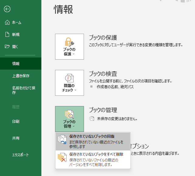
Excelが異常終了!開いていたExcelファイルを復元する方法 | システムエンジニアについてまじめに考えるブログ
Sold Shop & Retail Property At Pets Domain, 85 & 85A Wilson Street

Sold Shop & Retail Property at Pets Domain, 85 & 85A Wilson Street ...
About Us – Pets Domain

About Us – Pets Domain